


- Corneal Surgery

DMEK (Descemet’s Membrane Endothelial Keratoplasty)

- Treatment
- Corneal Surgery
DMEK (Descemet’s Membrane Endothelial Keratoplasty)
DMEK (Descemet’s Membrane Endothelial Keratoplasty) is a partial thickness cornea transplant that can restore lost vision when the endothelium, the innermost layer of cells of the cornea, are no longer functioning adequately.
What is a DMEK procedure ?
Descemet’s membrane endothelial keratoplasty (DMEK) is a corneal transplantation procedure, replacing only the innermost layers of the cornea, namely the Descemet membrane and endothelium, with human corneal donor tissue. This makes DMEK different from another partial thickness procedure, DSAEK ( Descemet’s stripping automated endothelial keratoplasty) which also replaces the posterior stroma. A DMEK procedure helps to improve corneal clarity against cloudiness caused by injury or other eye conditions like fuchs endothelial dystrophy.
DMEK procedure
The faulty inner layer of corneal cells is removed and replaced with the layer of cells taken from a donated human cornea. In a DMEK procedure the implanted donor material contains only Descemet’s membrane and corneal endothelial cells (15-20 microns thick) and is therefore a like-for-like replacement of tissue. Medical conditions including: Fuchs’ dystrophy, bullous keratopathy, iridocorneal endothelial (ICE) syndrome, or other endothelial disorders may cause blurry or cloudy vision and glare. An EK corneal transplantation selectively replaces only the diseased layer of the cornea, leaving healthy areas intact.


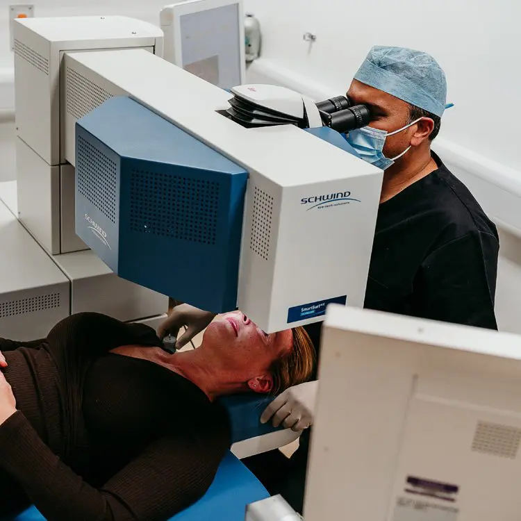
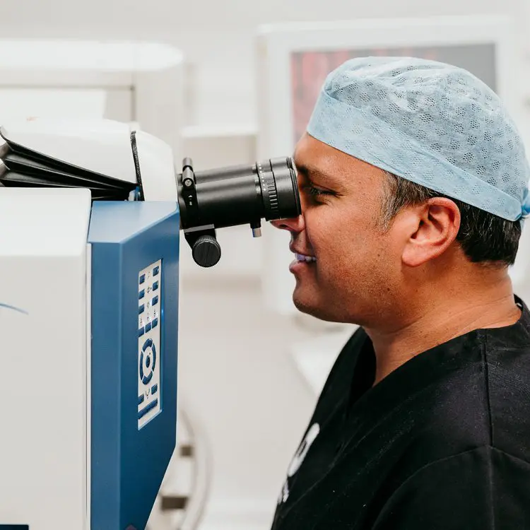
How is it performed?
- 1.Performed under local or general anaesthetic in sterile operating theatre.
- 2.Patient is laid flat on the treatment bed.
- 3.Povidone iodine clean, sterile drape applied, and eyelid support inserted.
- 4.Faulty / failing corneal endothelium is removed and the donor cornea endothelial transplant inserted.
- 5.Graft is secured in place with air or an SF6 bubble which tamponades the graft against the inside of the cornea.
- 6.Topical anti-inflammatory eye drops and antibiotic eye drops are applied and patients are advised to lie flat on their back as much as possible (45 minutes in every hour) for the first 2-3 days post-surgery.