


- Concerns

- Corneal

The cornea consists of six key layers. From front to back they include the epithelium, Bowman’s layer, the stroma, Dua’s layer, Descemet’s layer and the endothelium. Corneal dystrophies cause physiological changes to these layers which impair the optical performance of the eye. Two of the most common forms of corneal dystrophy aside from the aforementioned Fuchs dystrophy, include epithelial basement membrane dystrophy and lattice dystrophy.
What are the Symptoms?
What are the Causes?
Corneal dystrophies can be inherited through autosomal dominant and autosomal recessive inheritance patterns. However, in many cases a genetic link cannot be identified. Epithelial basement membrane dystrophy (otherwise known as map dot fingerprint or Cogan’s dystrophy) is caused by a compromised basement layer leaving the surface epithelium loose. This can cause uncomfortable erosions to form which require management by a corneal specialist.
Lattice dystrophy is acquired through a dominant inheritance pattern. Amyloid matter is displaced into the middle layer of the cornea resulting in a series of overlapping lines and dots which appear like a lattice.
What is the Treatment?
Corneal erosions arising from epithelial basement membrane dystrophy can be managed with regular daytime eye drops and the application of a viscous gel before sleep. Prophylactic antibiotic cover may also be prescribed to reduce the risk of infection. Lubrication should continue for 3 months to allow the epithelium to heal and bind. In the event of recurrent and challenging epithelial erosions, the surface skin can be debrided using the Scwhind Amaris 1050 RS laser. This procedure resets the epithelial surface and can provide effective long-term relief. Early stage lattice dystrophy can similarly be managed with lubrication. However, more advanced cases may require corneal grafts with our Laser Vision corneal specialists.

Treatment Options
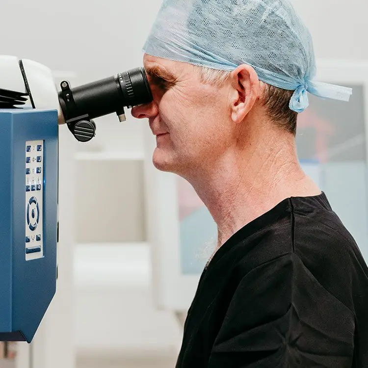
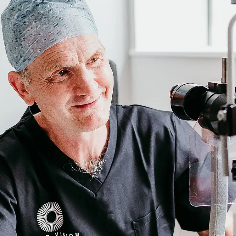
Choosing the right vision correction clinic for your surgery is paramount. This is a life changing procedure after all, and you need to have complete trust in your surgeon and care team of professionals.

More information about PTK
Phototherapeutic Keratectomy (PTK) at LaserVision is an established form of therapeutic laser eye surgery.
View PTKOur Technology
We invest in the latest equipment hand chosen by our surgeons, so that we can deliver outstanding results with the safest surgery possible.
0% APR FINANCE
We offer 0% APR* finance for up to 36 months. To find out more information about which treatments are available with 0% APR finance, contact us today.
*subject to status
*minimum age applies
*only available to UK residents aged 18 and over
At LaserVision, we focus on you. Each consultation, treatment, procedure and aftercare plan is specifically tailored to your needs and requirements so you can rest assured you are getting the best treatment for you.
Laser Vision Limited is authorised and regulated by the Financial Conduct Authority and is the broker and not the lender. Our FCA registration number is 805270. Laser Vision Limited offers credit products from Secure Trust Bank PLC trading as V12 Retail Finance. Credit is provided subject to affordability, age and status. Minimum spend applies. Not all products offered by Secure Trust Bank PLC are regulated by the Financial Conduct Authority
Please submit a compliment, concern, or complaint to: [email protected]. Every observation is an opportunity for our team to improve the quality of our patient care.
Sign up to our newsletter to receive updates
About
Our Patients
For Doctors
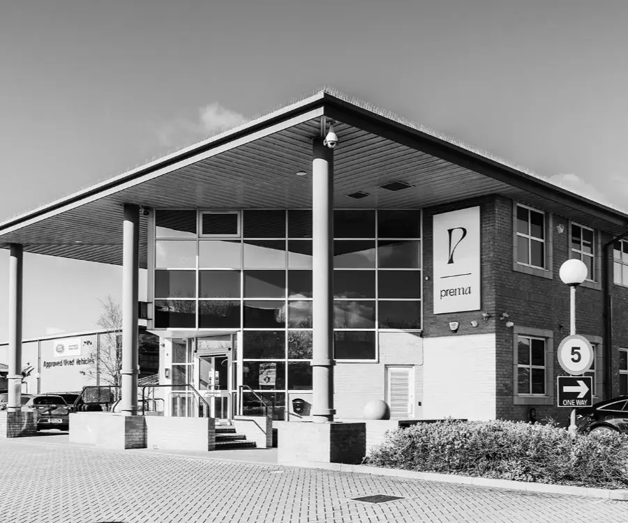
Office
LaserVision Ltd
Prema,
Compass Road
Portsmouth, PO6 4RP
United Kingdom
Company number 07313135
Contact Us
At LaserVision, we focus on you. Each consultation, treatment, procedure and aftercare plan is specifically tailored to your needs and requirements so you can rest assured you are getting the best treatment for you.
Laser Vision Limited is authorised and regulated by the Financial Conduct Authority and is the broker and not the lender. Our FCA registration number is 805270. Laser Vision Limited offers credit products from Secure Trust Bank PLC trading as V12 Retail Finance. Credit is provided subject to affordability, age and status. Minimum spend applies. Not all products offered by Secure Trust Bank PLC are regulated by the Financial Conduct Authority
Please submit a compliment, concern, or complaint to: [email protected]. Every observation is an opportunity for our team to improve the quality of our patient care.
Sign up to our newsletter to receive updates
![]() Site by Blow Media
Site by Blow Media