
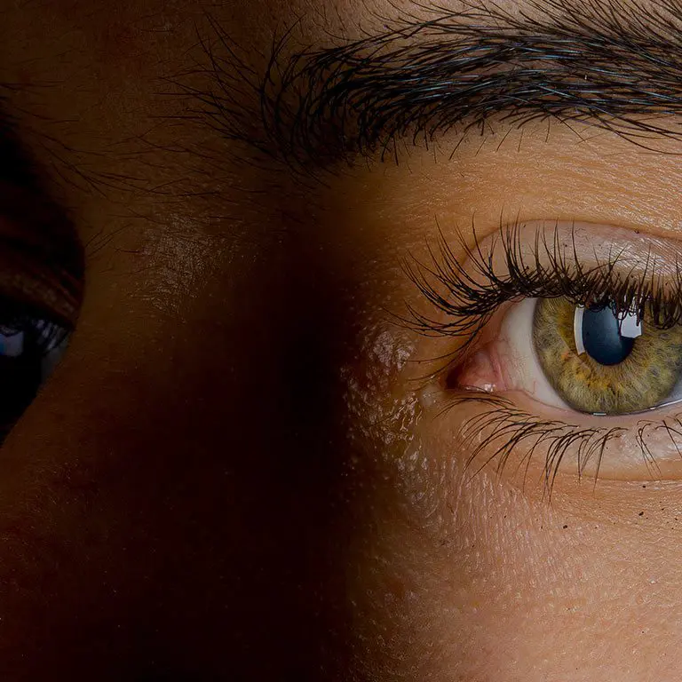


Central serous retinopathy (CSR) occurs when there is a build-up of fluid underneath the retina / macula.
It usually affects one eye and is self-limiting without treatment but may be recurrent in up to 50% of patients. It is six times more common in men than women, and most often affects people aged between the ages of 20 and 50.
A small percentage of patients develop a growth of abnormal vessels under the retina which leaks fluid and may require treatment with anti-VEGF injections. If chronic, the condition may damage the photosensitive cells of the retina leading to permanent visual loss.
What are the Symptoms?
What are the Causes?
The cause of CSR is not fully understood, however, it seems to run in families suggesting a genetic link. Patients with high blood pressure, heart disease or pregnancy have an increased risk of developing CSR.
Research also suggests other links for the cause of CSR, including: exposure to systemic steroids which can cause or worsen CSR, and patients with emotional distress and/or “type A” personalities.
What is the Diagnosis?
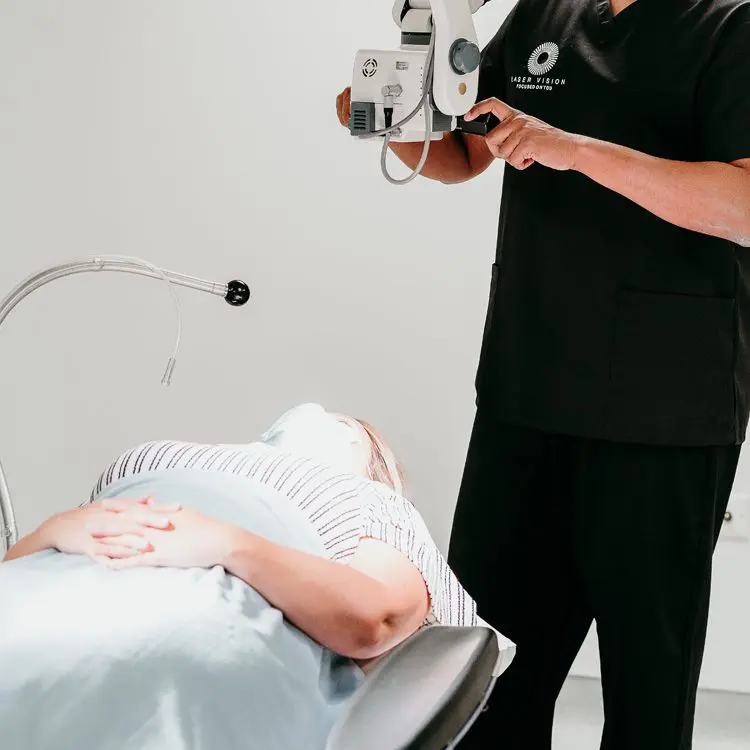
Central serous retinopathy (CSR) is diagnosed following a dilated retinal examination of the eye in combination with retinal imaging, using the following tests:
Optical Coherence Tomography (OCT) – an imaging test that uses light waves to take pictures of each of the retina’s layers, showing the presence of subretinal fluid and serous detachment.
Fluorescein angiography – a small amount of yellow dye (fluorescein) is injected into a vein in the arm and photographs taken of the back of the eye as the dye passes through the retinal vasculature.
The dye helps the ophthalmologist see how well blood is circulating inside the retina and any abnormal leakage from retinal blood vessels / under the retina. OCT angiography – combination of OCT retinal scanning and angiography gives higher resolution examination of the retinal structure and vasculature.


Treatment Options
Choosing the right vision correction clinic for your surgery is paramount. This is a life changing procedure after all, and you need to have complete trust in your surgeon and care team of professionals.
Our Technology
We invest in the latest equipment hand chosen by our surgeons, so that we can deliver outstanding results with the safest surgery possible.