


- Concerns

- Retina

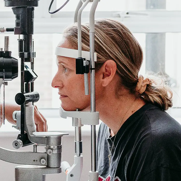
A retinal detachment is defined as the separation of neurosensory retina (light-detecting tissue) from the retinal pigment epithelium (base layer) at the back of the eye. Retinal detachment causes the retinal cells to separate from the blood vessels that provide oxygen and nourishment. If left untreated, retinal detachment can result in permanent loss of vision in the affected eye. Examination involves dilating the pupil with the assistance of the latest Optical Coherence Tomography (OCT) which is pivotal to managing this condition successfully.
What are the Symptoms?
What are the Causes?
There are three types of retinal detachment with their own precise pathophysiology: Rhegmatogenous (the most common form) – With this type of retinal detachment a break in the retinal tissue allows vitreous fluid to seep underneath the retina and detach it. Retinal tears and holes are far more common in high myopia (short-sighted prescriptions) as the retina is thinner and stretched. A posterior vitreous detachment (or PVD) in later life can also trigger a tear or hole;
Tractional – This is the result of scar tissue or the vitreous body tugging on the retina. Proliferative diabetic disease, trauma and surgery can all lead to scar tissue on the surface of the retina which can pull on the tissue to cause a defect;
Exudative – The formation of subretinal fluid due to inflammatory and/or exudative diseases including Sarcoidosis and metastatic cancers.


What is the Treatment?
If your retina detaches, it is urgent that you receive surgery to repair it as soon as possible – preferably within days of your diagnosis. There are a number of surgeries designed to seal holes and reattach the retina, your Laser Vision surgeon will recommend the best retinal detachment surgery for your case after a full consultation, including: Vitrectomy, Scleral Buckle or Pneumatic Retinopathy. In some cases a combination of Vitrectomy and Scleral Buckle are used.

Treatment Options
Choosing the right vision correction clinic for your surgery is paramount. This is a life changing procedure after all, and you need to have complete trust in your surgeon and care team of professionals.

More information about YAG Vitreolysis
YAG Vitreolysis is a non-invasive, pain-free laser procedure that can eliminate or greatly reduce the visual disturbance caused by floaters.
View YAG VitreolysisOur Technology
We invest in the latest equipment hand chosen by our surgeons, so that we can deliver outstanding results with the safest surgery possible.
0% APR FINANCE
We offer 0% APR* finance for up to 36 months. To find out more information about which treatments are available with 0% APR finance, contact us today.
*subject to status
*minimum age applies
*only available to UK residents aged 18 and over

At LaserVision, we focus on you. Each consultation, treatment, procedure and aftercare plan is specifically tailored to your needs and requirements so you can rest assured you are getting the best treatment for you.
Laser Vision Limited is authorised and regulated by the Financial Conduct Authority and is the broker and not the lender. Our FCA registration number is 805270. Laser Vision Limited offers credit products from Secure Trust Bank PLC trading as V12 Retail Finance. Credit is provided subject to affordability, age and status. Minimum spend applies. Not all products offered by Secure Trust Bank PLC are regulated by the Financial Conduct Authority
Please submit a compliment, concern, or complaint to: [email protected]. Every observation is an opportunity for our team to improve the quality of our patient care.
Sign up to our newsletter to receive updates
About
Our Patients
For Doctors
Office
LaserVision Ltd
Prema,
Compass Road
Portsmouth, PO6 4RP
United Kingdom
Company number 07313135
Contact Us
At LaserVision, we focus on you. Each consultation, treatment, procedure and aftercare plan is specifically tailored to your needs and requirements so you can rest assured you are getting the best treatment for you.
Laser Vision Limited is authorised and regulated by the Financial Conduct Authority and is the broker and not the lender. Our FCA registration number is 805270. Laser Vision Limited offers credit products from Secure Trust Bank PLC trading as V12 Retail Finance. Credit is provided subject to affordability, age and status. Minimum spend applies. Not all products offered by Secure Trust Bank PLC are regulated by the Financial Conduct Authority
Please submit a compliment, concern, or complaint to: [email protected]. Every observation is an opportunity for our team to improve the quality of our patient care.
Sign up to our newsletter to receive updates
![]() Site by Blow Media
Site by Blow Media