


1948 – Father Waclaw Szuniewicz – Changing Corneal Curvature
Polish missionary, ophthalmologist, pioneer of refractive surgery of the cornea.
PRINCIPLE CONTRIBUTIONS
He went to US in 1948. He continued his experimental work on surgical method on changing corneal curvature at Yale University in New Haven. He was the first to experiment with changing the shape of the cornea. His scientific accomplishments make him one of the pioneers of refractive surgery of cornea.
1964 – Professor Jos – Ignacio Barraquer – Introduction of Keratomileusis
PRINCIPLE CONTRIBUTIONS
After many years of continuous investigation into modification of the cornea (eg, corneal resections for the management of astigmatism, refractive lamellar grafts with and without freezing, the use of a cryolathe), two of Professor Jos – Ignacio Barraquer’s principle contributions to refractive keratoplasty occurred. In 1964, he introduced keratomileusis (ie, Greek for sculptured cornea)3 and an even more sophisticated technique, keratophakia (ie, Greek for corneal lens).
Keratomileusis. This is an autoplastic procedure for the correction of ametropia. The basis of the operation is to remove, modify, and reinsert the corneal disc into the patient’s eye. After fixation with a pneumatic fixation ring, a special microkeratome separates a parallel-faced corneal disk from the anterior layers of the cornea. The microkeratome operates on the principle of a carpenter’s plane, shaving the parallel-faced corneal disk, which is then frozen and carved with a special lathe according to computer measurements.
1970 – Svyatoslov N. Fyodorov, MD invents Radial Keratotomy
In the 1970s, Fyodorov saw a young myopic boy with corneal injuries caused by glass fragments. After the injury healed, Fyodorov found the boy’s vision was actually better than before the injury. He theorized the radial cuts flattened the cornea. Although a Japanese surgeon had already attempted refractive surgery by making incisions from within the eye, Fyodorov developed radial keratotomy by calculating how to make radial incisions on the anterior surface of the cornea to change its shape. His colleagues did not share his enthusiasm at first. Despite opposition, Fyodorov pushed forward with radial keratotomy.
1973-1983 – Invention of the Excimer Laser
Three researchers at the IBM ® Thomas J. Watson Research Center in Yorktown, New York – Samuel Blum, Rangaswamy Srinivasan and James J. Wynne – had been exploring new ways to use the excimer laser that had been recently acquired by their laser physics and chemistry group. Blum was an expert in materials science; Srinivasan was a photochemist with 21 US patents to his name; and Wynne was a physicist, who was the manager of the group at IBM. The excimer laser uses reactive gases, such as chlorine and fluorine, mixed with inert gases, such as argon, krypton and xenon. When electrically excited, the gas mixture emits energetic pulses of ultraviolet light, which can make very precise, minute changes to irradiated material, such as polymers.
1987 – Stephen Trokel, MD – First Used The Excimer Laser on the Cornea
Dr. Steven Trokel introduced Photorefractive Keratectomy (PRK). He also patented the Excimer laser for vision correction and performed the first laser surgery on a patient’s eyes in 1987.
Upon learning of the IBM work, ophthalmologist Stephen Trokel, affiliated with Columbia Presbyterian Medical Center in New York City, came to the Watson Research Center in the summer of 1983 to collaborate on experiments with Srinivasan and researcher Bodil Braren. Trokel, Srinivasan, and Braren wrote a paper introducing the idea of using the laser to reshape or sculpt the cornea (the clear covering on the front of the eye) in order to correct refractive errors, such as myopia or hyperopia. Their paper, published in a major ophthalmology journal in December 1983, launched a worldwide program of research to develop excimer laser-based refractive surgery.
New York City ophthalmologist, Steven Trokel made the connection to the cornea and performed the first laser surgery on a patient’s eyes in 1987. The next ten years were spent perfecting the equipment and the techniques used in laser eye surgery. In 1996, the first Excimer laser for ophthalmic refractive use was approved in the United States.
1991 – Stephen Slade MD / Stephen Brint – The First LASIK surgery
Dr. Stephen Slade and his scientific colleague, Dr. Stephen Brint, were the first ophthalmologists to perform LASIK in the United States.
The Advent of LASIK (Laser Assisted In Situ Keratomileusis)
With the development of precise surgical cutting instruments, the use of the excimer laser could be combined with an incision to produce a particular surgical result. It has become, by far, the most commonly performed refractive surgery procedure used today. During LASIK the surgeon first creates a thin corneal flap using a device called a microkeratome. The corneal flap is lifted up, and the excimer laser beam is applied to the exposed interior surface of the cornea to reshape the tissue. The flap is then replaced over the treated area. This corneal flap serves a natural bandage, which eliminates the discomfort associated with other types of refractive surgery, and expedites the healing process. Because of the extraordinary bonding properties of the corneal tissue, stitches are not needed to keep the flap in place after LASIK surgery.
1995 – PRK approved by FDA
PRK – Photorefractive Keratectomy
Photorefractive keratectomy, or PRK, was the first kind of corrective eye surgery to use a laser rather than a blade to remove corneal tissue. Though the excimer laser was developed in the early 1970s and modified for ophthalmic use in the early 1980s, the Food and Drug Administration did not approve its use for PRK corrective eye surgery until 1995. PRK was originally developed to treat myopia by removing a small amount of the cornea with a laser. Later refinements in the technology allowed surgeons to also treat patients with hyperopia, or farsightedness, and astigmatism. Unlike the previous technique of ALK (and LASIK, which had not yet been developed), PRK does not require the creation of a flap in the cornea. Instead, the laser is applied directly to the surface of the cornea. As a result, recovery from PRK corrective eye surgery is rather lengthy and uncomfortable. PRK became less and less popular following the development of LASIK, a procedure that allowed patients to have their vision corrected without the need for extended recovery from surgery. In fact, only ten years after its initial FDA approval, PRK corrective eye surgery was performed in only a small percentage of surgical eye correction treatments.
1996 – LASIK approved by FDA
The idea of combining previously proven flap technology and reshaping the cornea with the excimer laser occurred in the early 1990s and the dawn of LASIK surgery began. LASIK surgery was initially performed throughout the 1990s in the US as an”off-label” use of the excimer laser. The initial clinical trials for LASIK began in 1996.
2001 – Evolution of Femtosecond Laser in Refractive Surgery
In 1997, Dr Tibor Juhasz, a biomedical engineer familiar with earlier femtosecond research began pondering over medical applications. The first femtosecond laser approved for bladeless LASIK in the United States was the IntraLase laser, which gained FDA approval in 2001. IntraLase Inc. later introduced several new models of this laser with advanced features.
2003 – Wavefront Technology Approved
RAISING THE BAR: CUSTOMIZED WAVEFRONT LASIK
In May, 2003, the FDA approved the use of the Customized Wavefront LASIK using the VISX CustomVue System, an entirely new generation of laser vision correction and the predecessor of the system used by Dr. Shapiro. With CustomVue custom wavefront LASIK, the goal of surgery is no longer to equal the best glasses or contacts, but to actually exceed the quality of vision of glasses or contacts. The results of the FDA trials were outstanding.
Request a Brochure

0% APR FINANCE
We offer 0% APR* finance for up to 36 months. To find out more information about which treatments are available with 0% APR finance, contact us today.
*subject to status
*minimum age applies
*only available to UK residents aged 18 and over

At LaserVision, we focus on you. Each consultation, treatment, procedure and aftercare plan is specifically tailored to your needs and requirements so you can rest assured you are getting the best treatment for you.
Laser Vision Limited is authorised and regulated by the Financial Conduct Authority and is the broker and not the lender. Our FCA registration number is 805270. Laser Vision Limited offers credit products from Secure Trust Bank PLC trading as V12 Retail Finance. Credit is provided subject to affordability, age and status. Minimum spend applies. Not all products offered by Secure Trust Bank PLC are regulated by the Financial Conduct Authority
Please submit a compliment, concern, or complaint to: [email protected]. Every observation is an opportunity for our team to improve the quality of our patient care.
Sign up to our newsletter to receive updates
About
Our Patients
For Doctors
Office
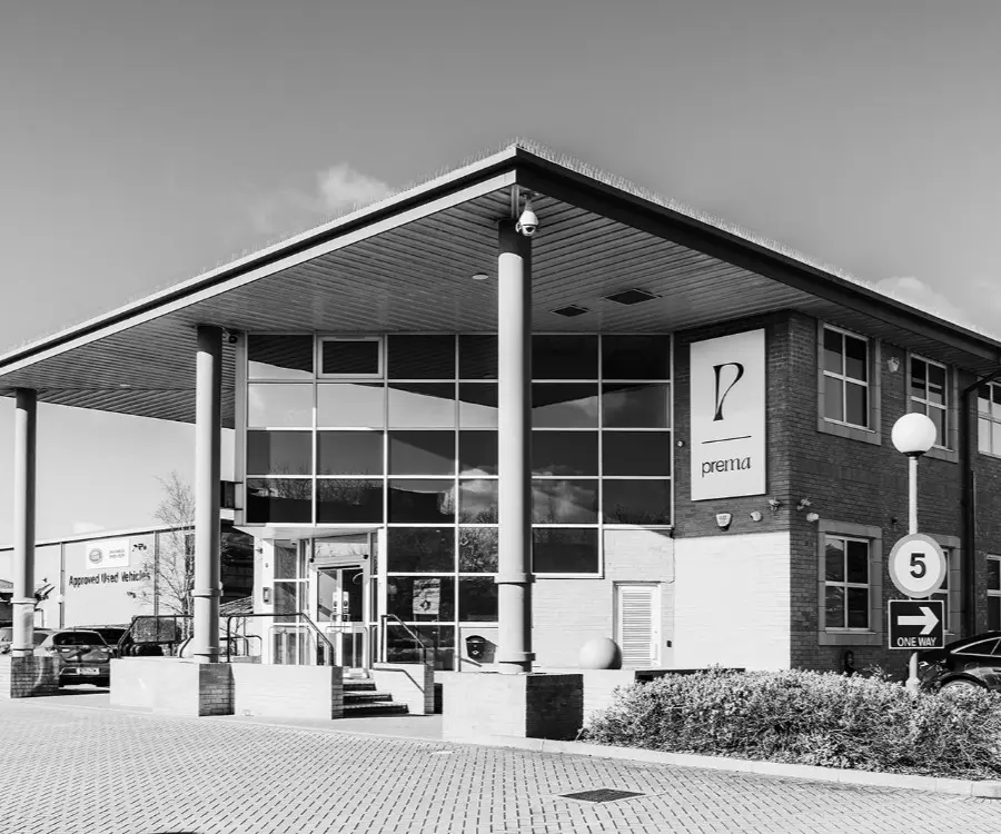
LaserVision Ltd
Prema,
Compass Road
Portsmouth, PO6 4RP
United Kingdom
Company number 07313135
Contact Us
At LaserVision, we focus on you. Each consultation, treatment, procedure and aftercare plan is specifically tailored to your needs and requirements so you can rest assured you are getting the best treatment for you.
Laser Vision Limited is authorised and regulated by the Financial Conduct Authority and is the broker and not the lender. Our FCA registration number is 805270. Laser Vision Limited offers credit products from Secure Trust Bank PLC trading as V12 Retail Finance. Credit is provided subject to affordability, age and status. Minimum spend applies. Not all products offered by Secure Trust Bank PLC are regulated by the Financial Conduct Authority
Please submit a compliment, concern, or complaint to: [email protected]. Every observation is an opportunity for our team to improve the quality of our patient care.
Sign up to our newsletter to receive updates
- © Copyright 2026
- Privacy
- Complaints
- Modern Slavery
- Sitemap
- Carbon Reduction Plan
![]() Site by Blow Media
Site by Blow Media